More accurate diagnostics through natural weight bearing and head movement.
Have a more meaningful examination while sitting or standing.
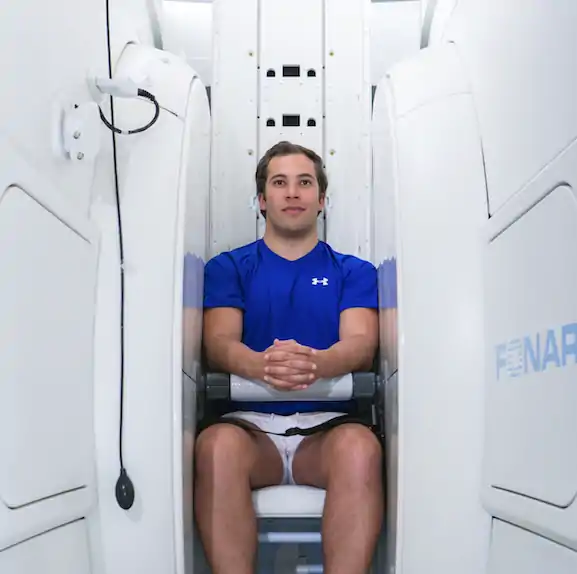
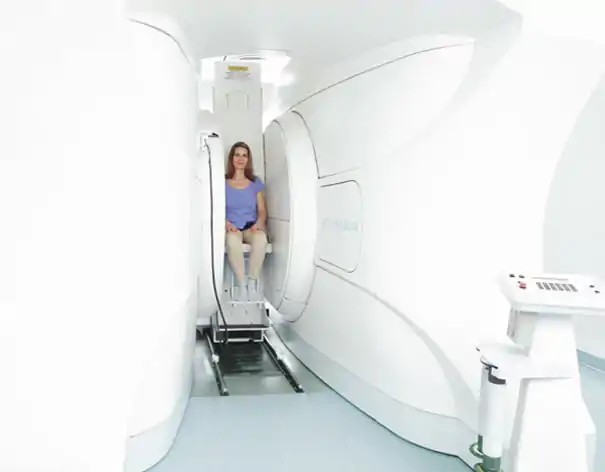
Fully open construction offers a much higher level of comfort with freedom of view & volume.
No constricting tubes or stifling “sandwiches”!
A better view of a higher quality of life

Recognise true cause
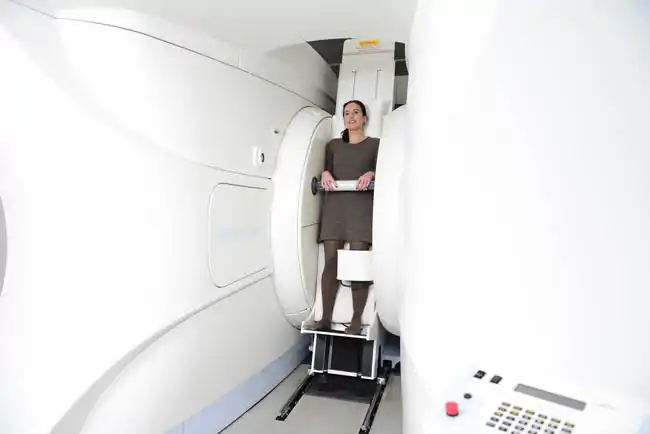
Causes of pain and triggers of complaints finally become visible. Why? Because the upright MRI, also called sitting MRI, enables the examination of the very complex upper cervical joint and cervical vertebra region in different head positions, e.g. also lateral inclinations. The examination is performed upright and under the natural weight load – which is impossible in conventional systems. Only the completely open design of the upright MRI allows magnetic resonance imaging examinations in which the upper cervical region can be examined in detail in different positions and thus provides more meaningful diagnostic results.
Sit down relaxed
Compared to the narrow tube systems, the sitting MRI offers a much higher patient comfort in terms of freedom of movement and view as well as volume. You have a free view out of the system during the examination and look at a large monitor on which you can follow the current TV programme or watch video films. It is also usually not necessary to wear headphones – as is the case with closed MRI systems – because the Upright MRI works very quietly. It is even possible for a person you trust to accompany you into the MRI room.


The examination procedure
Before every examination, there is a detailed discussion with the radiologist, during which please do not hesitate to ask any questions that are close to your heart. Only then does the actual examination begin, during which you will not be left alone at any time. To do this, you simply sit or stand in the system, depending on the position in which you are to be examined. While you take a short seat in the rest area after the examination, the images taken of you are analysed and the findings are drawn up. The radiologist will then discuss these findings with you in detail on the basis of the images.
